Continue
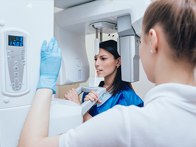
Low radiation imagery utilizes computer technology and digital sensors for the acquisition, viewing, storage, and sharing of diagnostic radiographic images. It offers several advantages over the older traditional film-based methods of taking x-rays. The most significant is that it reduces a patient’s exposure to x-ray radiation. This highly advanced technology also means that critical diagnostic images can be viewed instantly after being taken and easily shared and discussed with patients as well as other specialists or offices as required. Low radiation imagery is also safer for the environment as it does not require any chemicals or paper to develop.
An electronic pad, known as a sensor, is used instead of film to acquire a digital image. After the image is taken, it goes directly into the patient’s file on the computer. Once it is stored on the computer, it can be easily viewed on a screen, shared, or printed out.